







 Book an appointment
VÒNG TRÁNH THAI GÂY THỦNG THÀNH TRỰC TRÀNG
 Posted date: 14/01/2026
 Posted by: Nguyễn Kim Bình

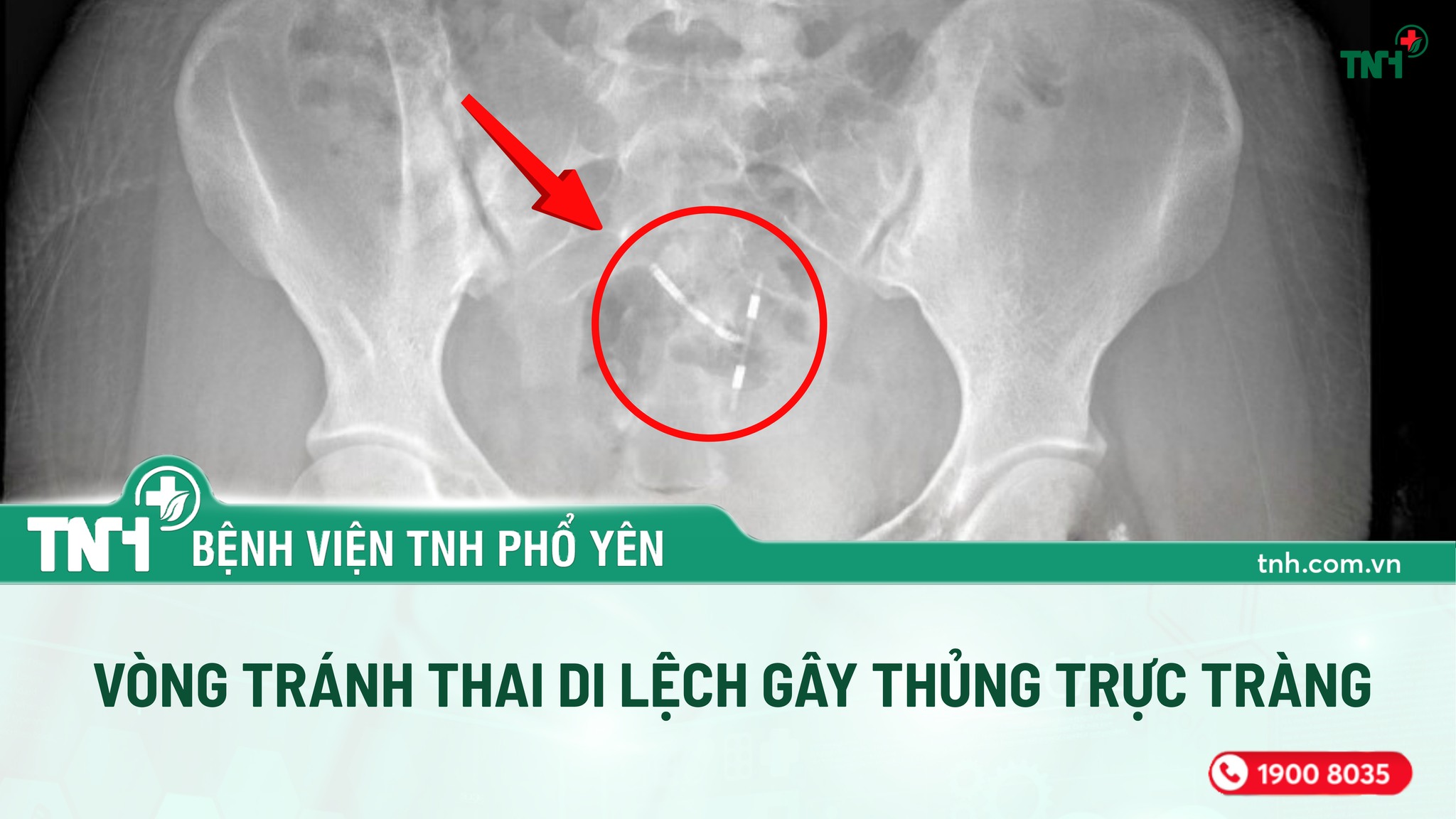
The Functional Diagnostics Department – TNH Pho Yen Hospital (Thai Nguyen) has recently managed an extremely rare clinical case: a T-shaped intrauterine device (IUD) migrated from its original position, perforated the rectal wall, causing inflammation and the formation of an intra-abdominal abscess.
A 21-year-old female patient was admitted with persistent abdominal pain. Her general condition was relatively stable, with no fever or vomiting. On clinical examination, doctors noted tenderness around the right periumbilical area, marked pain at McBurney’s point, and no signs of generalized peritonitis. With these initial symptoms, the condition could easily be mistaken for acute appendicitis or gynecological disorders.
Initial laboratory tests showed elevated white blood cell count and increased C-reactive protein (CRP), indicating an inflammatory process. However, abdominal ultrasound did not reveal any typical findings or clearly identify organ damage. Faced with a misleading clinical presentation and inconclusive imaging results, the medical team at TNH Pho Yen Hospital carefully reassessed the entire clinical course and decided to perform a colonoscopy to accurately determine the cause of the patient’s prolonged abdominal pain.
During the colonoscopy at the Functional Diagnostics Department, the doctors made a surprising discovery: an intrauterine device was located in the rectum. The stem of the device had perforated the rectal wall, while the transverse arms lay within the abdominal cavity. At the perforation site, inflammatory granulation tissue and purulent discharge were observed, indicating an ongoing infection. This was a dangerous situation because the rectum contains a large number of bacteria; damage to the intestinal wall significantly increases the risk of widespread infection, pelvic abscess formation, or even generalized peritonitis if the foreign body continues to migrate or is not promptly treated.
According to the doctors, intrauterine devices are a common and relatively safe method of contraception when properly placed within the uterine cavity. However, in rare cases, an IUD may migrate, perforate the uterine wall, and move to adjacent organs such as the bladder, abdominal cavity, or intestines. An IUD perforating the rectal wall and remaining there long enough to cause inflammation and abscess formation is extremely uncommon in clinical practice, making the initial diagnosis particularly challenging.
After an urgent consultation and assessment of the extent of injury, the medical team decided to intervene by removing the foreign body via colonoscopy. This was considered the optimal approach, as the patient showed no signs of generalized peritonitis and her overall condition was stable.
The procedure was performed using specialized endoscopic forceps. The doctors carefully stabilized the foreign body and gently removed the IUD from the rectal wall with precision, minimizing the risk of further tissue damage. The greatest challenge of this case was that the device had already perforated the rectal wall and was surrounded by inflamed tissue and an abscess. Any inaccurate maneuver could have worsened the injury, leading to bleeding or necessitating open surgery. However, thanks to the team’s expertise, close coordination, and meticulous technique, the foreign body was successfully removed without complications during or after the procedure, sparing the patient from major surgery.
Dr. Dinh Van Hung, Specialist Level I, Head of the Functional Diagnostics Department, stated: “This is a very rare case with clinical manifestations that can easily be confused with other surgical or obstetric emergencies. Accurate diagnosis and timely intervention helped the patient avoid severe complications and major surgery.”
After the procedure, the patient continued to receive anti-inflammatory treatment according to protocol and recovered well. Her abdominal pain significantly decreased, bowel movements returned to normal, and her health is now stable. She has since been discharged from the hospital.
Although IUDs are a safe and effective contraceptive method widely used, they still carry a risk of migration or uterine perforation in rare cases, especially in patients who do not undergo regular follow-up examinations or who have anatomical abnormalities or a history of gynecological interventions. Doctors emphasize the importance of regular follow-up after IUD insertion: attending scheduled check-ups, undergoing ultrasound to confirm proper device positioning, and promptly seeking medical attention when experiencing unusual abdominal pain are key factors in early detection and timely management of complications.
At the same time, this case clearly demonstrates the professional expertise and strong sense of responsibility of the medical staff at TNH Pho Yen Hospital in successfully managing rare and complex cases, contributing to patients’ peace of mind and trust when choosing healthcare services at the hospital.