







 Book an appointment
PULMONARY ARTERIOVENOUS MALFORMATION DETECTED IN A 34-YEAR-OLD FEMALE PATIENT
 Posted date: 17/09/2026
 Posted by: Nông Đức Thắng


Quick view of main content
An abnormality detected from a pulmonary opacity
What is a pulmonary arteriovenous malformation?
The role of diagnostic imaging in detecting PAVMs
Do not overlook abnormalities detected incidentally
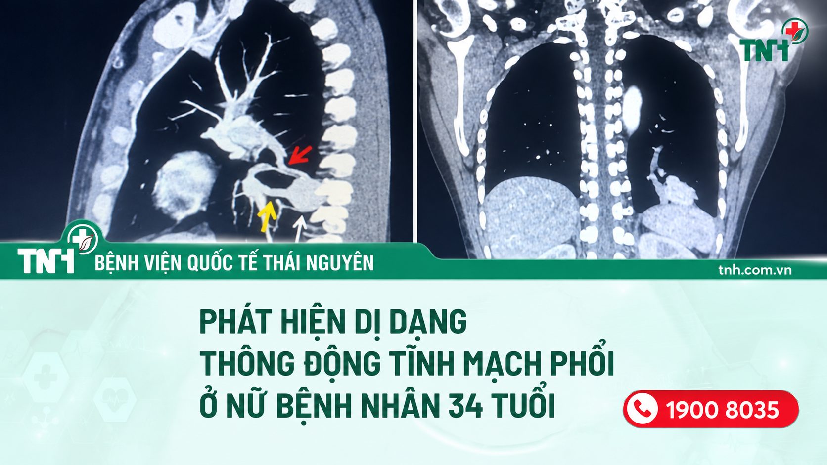
A 34-year-old female patient with no symptoms of cough, fever, or shortness of breath was diagnosed with a pulmonary arteriovenous malformation (PAVM) during an examination at Thai Nguyen International Hospital. This case highlights that certain pulmonary vascular abnormalities may remain asymptomatic for a long time and be detected incidentally through diagnostic imaging.
An abnormality detected from a pulmonary opacity
Previously, during a health check-up at another medical facility, the patient was found to have an opacity at the base of the left lung. She then visited Thai Nguyen International Hospital for further examination and evaluation.
At the time of examination, the patient had no cough, fever, or shortness of breath, and her general condition was stable. After clinical examination and medical history assessment, the doctors ordered a chest X-ray and contrast-enhanced chest CT scan.
Diagnostic imaging revealed that the lesion was vascular in origin. Based on its characteristics, location, and associated blood vessels, the doctors diagnosed the patient with a pulmonary arteriovenous malformation (PAVM).
What is a pulmonary arteriovenous malformation?
A pulmonary arteriovenous malformation is an abnormal connection between a pulmonary artery and a pulmonary vein, allowing blood to flow directly from the artery to the vein without passing through the normal pulmonary capillary network.
This abnormal connection may cause some blood to bypass normal oxygen exchange before returning to the systemic circulation. It may also increase the risk of complications related to embolism or blood vessel rupture.
PAVMs may remain asymptomatic for a long period, and many patients experience no obvious symptoms. In some cases, the condition is discovered incidentally on a chest X-ray or CT scan performed for another reason.
When symptoms do occur, patients may experience shortness of breath on exertion, fatigue, dizziness, cyanosis, cough, or recurrent nosebleeds. Importantly, PAVMs may be associated with serious complications such as stroke, brain abscess, and certain cardiovascular complications.
The role of diagnostic imaging in detecting PAVMs
A chest X-ray can help identify abnormalities in the lungs and may prompt doctors to recommend further diagnostic evaluation.
Meanwhile, contrast-enhanced chest CT provides a more detailed assessment of vascular lesions, including the location and size of the malformation, as well as the feeding artery and draining vein. This information is important for evaluating the patient's condition and determining an appropriate monitoring and management plan.
Do not overlook abnormalities detected incidentally
The case of this 34-year-old female patient at Thai Nguyen International Hospital demonstrates that certain pulmonary abnormalities may exist silently even when a person has no noticeable symptoms.
Therefore, if an abnormality is detected on a chest X-ray or during a routine health check-up, patients should seek medical evaluation and undergo further examinations as recommended by their doctor. Early detection, accurate diagnosis, and appropriate monitoring and management can help reduce the risk of serious complications.
TNH HOSPITAL GROUP JOINT STOCK COMPANY
Hotline: 1900 8035
Website: tnh.com.vn
Hospital Network:
Thai Nguyen International Hospital – 328 Luong Ngoc Quyen Street, Phan Dinh Phung Ward, Thai Nguyen
TNH Pho Yen Hospital – Tran Nguyen Han Street, Truong Thinh Residential Group, Van Xuan Ward, Thai Nguyen
TNH Viet Yen Hospital – Nguyen The Nho Street, Viet Yen Ward, Bac Ninh
TNH Lang Son Hospital – Block 10, Nhi Thanh Street, Tam Thanh Ward, Lang Son