







 Book an appointment
HYPERECHOIC BREAST MASSES ON ULTRASOUND ARE NOT ALWAYS BENIGN
 Posted date: 25/06/2026
 Posted by: Nông Đức Thắng

Quick view of main content
What Is a Hyperechoic Lesion on Ultrasound?
Common Benign Hyperechoic Breast Lesions
When Can a Hyperechoic Lesion Be Malignant?
What Additional Factors Will Physicians Evaluate?
Early Evaluation Is Key to Breast Health
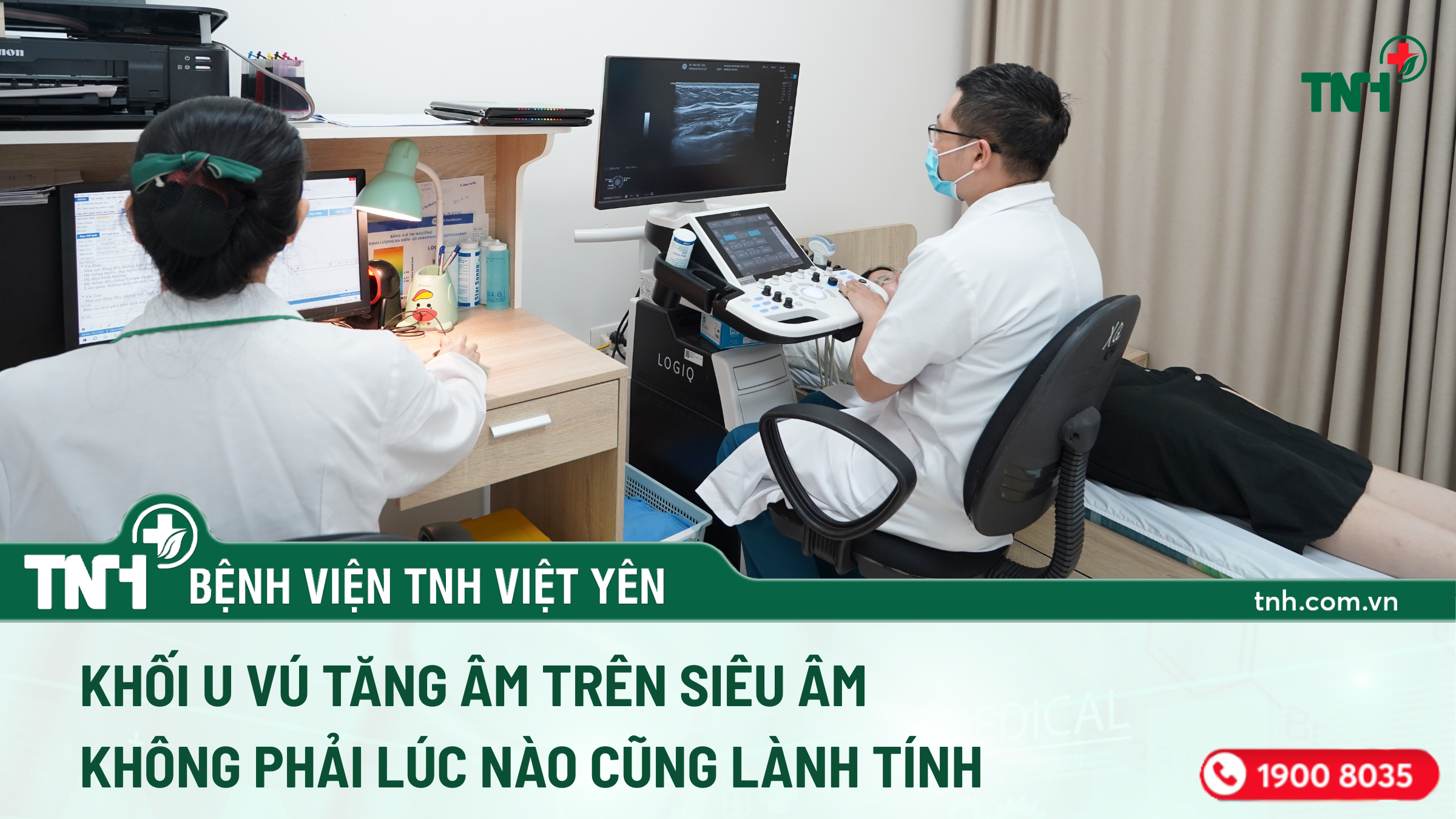
Many people assume that when a breast ultrasound reveals a “hyperechoic” lesion, there is no need for concern because it is considered a sign of a benign mass. However, in reality, not all hyperechoic lesions are harmless. Determining the true nature of a breast mass requires a comprehensive evaluation to avoid overlooking potentially malignant conditions.
What Is a Hyperechoic Lesion on Ultrasound?
On ultrasound imaging, a hyperechoic lesion appears brighter than the surrounding breast tissue because it reflects more ultrasound waves. While the majority of hyperechoic breast lesions are benign, certain malignant breast diseases may also present with hyperechoic characteristics.
Common Benign Hyperechoic Breast Lesions
Several benign breast conditions may appear hyperechoic on ultrasound, including:
- Hematoma following trauma or surgery
- Breast abscess and mastitis
- Fat necrosis
- Hamartoma
- Lactating adenoma in pregnant or breastfeeding women
- Hemangioma and angiolipoma
- Other benign breast tumors
These conditions generally have a favorable prognosis and can be managed through appropriate monitoring or treatment, depending on the individual case.
When Can a Hyperechoic Lesion Be Malignant?
Although less common, some malignant breast tumors may also appear hyperechoic on ultrasound, including:
- Invasive ductal carcinoma
- Invasive lobular carcinoma
- Breast lymphoma
- Angiosarcoma
- Certain rare types of breast sarcoma
Therefore, the presence of a hyperechoic lesion alone is not sufficient to determine whether a breast mass is benign or malignant.
What Additional Factors Will Physicians Evaluate?
To establish an accurate diagnosis, physicians consider multiple clinical and imaging factors, such as:
- Shape of the lesion
- Margin characteristics
- Internal vascularity
- Size and changes over time
- Clinical examination findings
- Mammography or breast MRI when indicated
- Biopsy for definitive pathological diagnosis
Early Evaluation Is Key to Breast Health
The detection of a hyperechoic lesion on breast ultrasound does not automatically indicate a benign condition. Each case should be thoroughly assessed by a specialist to determine the most appropriate follow-up or treatment plan.
If you notice any breast abnormalities, such as a palpable lump, persistent pain, skin changes, or abnormal nipple discharge, seek medical evaluation promptly for accurate diagnosis and timely management.
Key Message:
“A hyperechoic breast lesion does not necessarily mean a benign lesion.”
For further consultation and support, please contact our Hotline at 1900 8035 or send a message via our Fanpage.